Medical imaging is a fundamental part of health diagnosis and management. From X-ray to ultrasound, MRI, CT scan, and PET scan, the range of imaging technologies available grants physicians an unparalleled ability to observe the human body without requiring surgery.
Every year, an astonishing 3.6 billion medical diagnostics are conducted around the globe. To ensure that people receive quality and appropriate healthcare, it is vital to comprehend the various forms of medical imaging accessible so we can make educated decisions about our health.

Overview of Medical Imaging Technologies and Their Uses
Thanks to the emergence of medical imaging solutions, physicians now have an unprecedented level of accuracy and effectiveness when diagnosing and treating their patients. From X-rays to MRIs, each imaging technology is designed for a specific purpose and delivers valuable insights that doctors can use. With this wide range of options at their fingertips, healthcare professionals are more capable than ever before of uncovering health problems while providing efficient treatments.
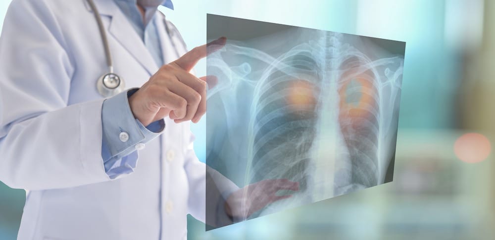
X-Rays
X-rays are a revolutionary medical technology that has drastically changed the way doctors diagnose bone fractures, illnesses, and other medical issues in our bodies. The power of this incredible science is simply awe-inspiring. Since the first discovery of this miraculous invention more than a hundred years ago, trained professionals have benefited from its usefulness.
By transmitting high-energy electromagnetic waves through the body, X-rays create 2D images on film that are invaluable in helping to identify broken bones and sometimes even detect cancerous tumors. Utilizing this powerful technology empowers doctors with the capacity for more precise diagnoses—one of its most essential functions.
CAT Scans
Medical professionals everywhere have embraced CAT scans to accurately diagnose a range of conditions. But how does a CAT scan work, exactly? Essentially, a patient lies down on a table that moves through a tube-like machine. This machine then sends out a series of X-rays that are picked up by detectors and converted into images. These images can be incredibly helpful in spotting the signs of various conditions, such as tumors, fractures, and more.
Ultrasound Images
Ultrasound imaging has revolutionized the medical field, offering physicians an unprecedented ability to observe internal organs and tissues in high detail. This has drastically improved the diagnostic accuracy of conditions such as cancer, heart disease, pregnancy complications, and more. Thanks to advances in ultrasound technology we can now identify potential issues far more quickly than ever before.
Ultrasound imaging is an invaluable tool around the world, offering countless advantages that make it a preferred diagnostic method. The non-invasive approach eliminates painful procedures and radiation exposure while also providing cost efficiency and portability. With its wide availability in medical settings worldwide, ultrasound images are among the most reliable ways to monitor patient health quickly and accurately.

MRI Scans
An MRI scan, short for magnetic resonance imaging, works by using a strong magnetic field and radio waves to produce images of our internal structures. As soon as you step into the MRI machine, its strong magnetic field causes your body’s hydrogen atoms to line up. Then, radio waves pass through your body and cause these particles to give off signals that sensors in the system detect.
These signals are then processed by a computer into detailed, cross-sectional images of your body. With MRI technology, doctors can diagnose a range of conditions, from torn ligaments to brain tumors, and provide the best treatment options for their patients.
PET Scans
PET scans are an incredibly useful tool in detecting cancerous cells. The secret to their success lies in the use of radioactive substances. These substances, known as radiotracers, are injected or ingested into the body and then absorbed by cells.
Cancer cells have a higher metabolic rate than healthy cells, meaning they absorb more radiotracers. This increased absorption shows up as bright spots on the PET scan, allowing doctors to identify the cancerous cells and determine the extent of the disease. While the use of radioactive substances can be intimidating, the benefits of early cancer detection far outweigh any potential risks.
In recent years, research into VHH antibodies, also known as nanobodies, has shown promise in enhancing PET scan technology. Derived from camelid immune systems, these small, single-domain antibodies exhibit exceptional stability and high specificity for disease biomarkers.This specificity is being explored to develop radiotracers that can improve the accuracy of PET imaging. Early studies suggest that VHH antibody-based radiotracers may provide clearer images and more precise diagnostics, particularly for detecting and monitoring cancer and other conditions.
Bottom Line
Health professionals rely on medical imaging to detect and track a range of conditions. When patients gain an understanding of the various forms of medical imaging services, they can make more informed decisions about their health and ensure that they are receiving top-notch care. Thanks to modern technology, we have access to incredibly detailed views inside our bodies, helping us acquire better insight into not only physical health but also overall well-being.

The Editorial Team at Healthcare Business Today is made up of experienced healthcare writers and editors, led by managing editor Daniel Casciato, who has over 25 years of experience in healthcare journalism. Since 1998, our team has delivered trusted, high-quality health and wellness content across numerous platforms.
Disclaimer: The content on this site is for general informational purposes only and is not intended as medical, legal, or financial advice. No content published here should be construed as a substitute for professional advice, diagnosis, or treatment. Always consult with a qualified healthcare or legal professional regarding your specific needs.
See our full disclaimer for more details.