Finding out that you or your loved ones have been diagnosed with cancer is one of the most stressful moments to go through. In such cases, it cannot be easy to stay optimistic. If it’s at an advanced stage, it becomes even more difficult to handle it.
One major type of cancer that has been on the rise and affects more people due to factors such as lifestyle is lung cancer. This type of cancer can be severe, especially if diagnosed later, as it cannot be cured by surgery and other therapeutic methods. As a result, there has been a great demand for new ways to diagnose and treat lung cancer at an early stage.
Thanks to technology, the diagnosis of lung cancer at an early stage are increasing, and it has helped with the treatment procedures. While some of these technologies can be used individually, a combination of different sets is required in some cases to ensure accuracy.
Here are some of the said technologies:
- Intraoperative CT Imaging
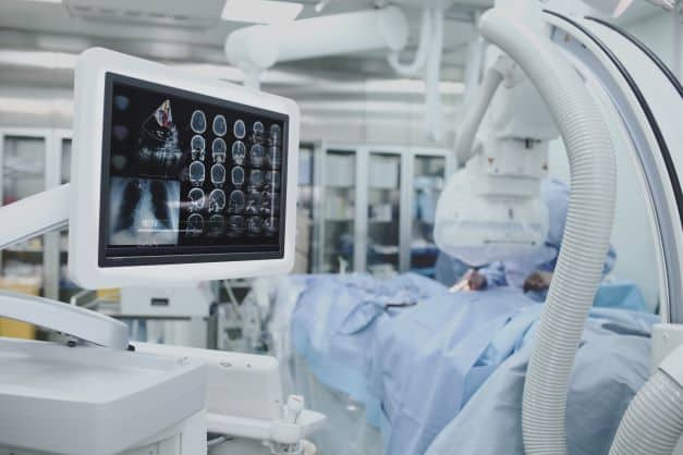
Body Vision Medical’s (https://bodyvisionmedical.com) intraoperative CT imaging system allows pulmonologists to visualize the actual lung lesion and lesion location during a diagnostic procedure using any conventional C-arm, a standard piece of x-ray equipment found in virtually every bronchoscopy suite. This means physicians can confidently biopsy smaller, more difficult-to-access lung lesions at earlier stages. This drastically increases the chances of an early, definitive diagnosis for potential lung cancer patients, improving the probability of timely treatment and patient survival.
The Body Vision system seamlessly integrates with existing tools and equipment, including robotic bronchoscopy platforms, making it simple for pulmonologists to integrate this advanced imaging technology into their practice.
This method is a safe and accurate procedure in evaluating patients with pulmonary nodules. No other technology readily accessible to pulmonologists enables this kind of image-guided biopsy that has been clinically proven to maximize the likelihood of obtaining a conclusive diagnosis.
Chest computed tomography (CT)
Chest CT is an imaging tool used as a noninvasive modality for screening and staging lung cancer. It’s fundamental in evaluating lung cancer as it helps define the size, location, and characteristics of lung lesions. Low-dose chest CT has been used to screen cancer in high-risk individuals. This is effective in detecting small lung nodules compared to conventional chest radiography. (2)

A trial demonstration by the US National Lung Cancer Screening Trial shows that using low dose Chest CT to screen individuals with high risk has helped reduce associated mortality by 20%. Even though the patients with positive results need further diagnosis using other procedures, this technology has gone to help with screening and diagnosing lung cancer. (2)
- Brain magnetic resonance imaging
Lung cancer is one of the significant causes of brain metastases. So, conducting brain magnetic resonance imaging is a great way to detect lung cancer in individuals. Brain metastases can be detected as an early symptom, and their presence is essential when considering the therapeutic agents and treatment modality to be used on the patient. (2)
Also, the Hazard Identification and Risk Authority (HIRA) guidelines recommend that brain magnetic resonance imaging be used for the initial screening and staging of lung cancer. This is also backed up by the fact that brain metastases are caused mainly by lung cancer, so screening and staging them play a significant role in the diagnosis. (2)
- Artificial intelligence (AI) analysis
Using AI-trained algorithms is also an excellent way to detect pulmonary nodules and improve lung cancer detection on chest radiographs. This is according to a study that tests the sensitivity and specificity in finding malignant pulmonary nodules. The result of this study was 94% and 83%, respectively. AI has also shown to have a higher sensitivity than the National Lung Screening Trial radiologists, which indicates it has a higher possibility of detecting lung cancer. As a result, it can be used as a second reader. (3)
Moreover, AI bases its detection procedures on deep learning. This is done by looking at real-life examples of tumors instead of a programmer’s definition of tumors. Researches feed the system with large data sets. From this data, the system will keep learning and improving over time. Therefore, AI can reliably distinguish between tumors and splotches. They can also help clinicians make informed decisions regarding the treatment procedure. (4)
Conclusion
Lung cancer is one of the most prevalent and deadliest types of cancer today. There have been cases of people succumbing to it due to failure to detect the cases at an early stage when it can be controlled. This means that when cases are detected early enough, they can be managed.
Conventional methods of diagnosing failed to detect the early stages of lung cancer, leading to a high mortality rate. However, as the demand for early diagnosis increased, new and improved technology, such as C-arm-based tomography and AI, has improved detection.
References
- “C-Arm Cone-Beam CT-Guided Transthoracic Lung Core Needle Biopsy as a Standard Diagnostic Tool: An Observational Study”, Source: https://www.researchgate.net/publication/274259169_C-Arm_Cone-Beam_CT-Guided_Transthoracic_Lung_Core_Needle_Biopsy_as_a_Standard_Diagnostic_Tool_An_Observational_Study
- “Recent advances in diagnostic technologies in lung cancer”, Source: https://www.ncbi.nlm.nih.gov/pmc/articles/PMC7060993/
- “AI Analysis Can Improve Lung Cancer Detection on Chest Radiographs”, Source: https://www.itnonline.com/content/ai-analysis-can-improve-lung-cancer-detection-chest-radiographs
- “Artificial intelligence is improving the detection of lung cancer”, Source: https://www.nature.com/articles/d41586-020-03157-9