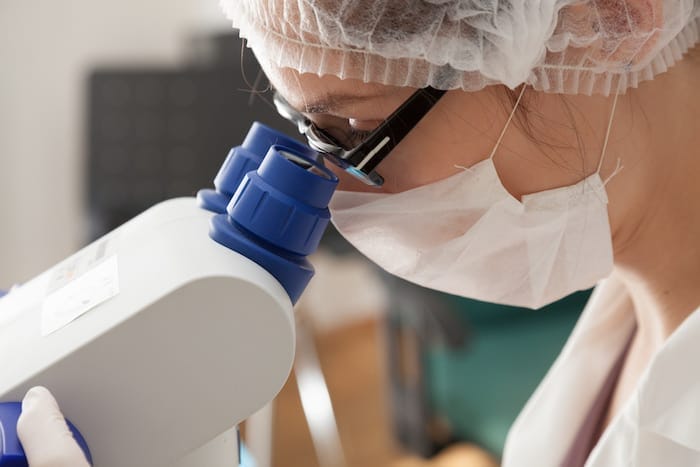
A polarizing microscope is simply a basic microscope added with accessories and polarizing filters. This type of ensemble allows users to examine attributes of double refracting substances like crystals. At first, an expert invented the polarizing microscope to help geologists assess crystalline patterns in minerals and rocks.
These days, this particular microscope has gone a long way and now plays a vital role in other areas like the industrial sector. The microscope helps in distinguishing asbestos, chemicals, fibers, and other materials. It can likewise recognize errors in semi-conductors and look for pressure points in glasses and metals.
Additionally, other disciplines such as biological and medical fields now also employ the improved polarizing microscope for testing, assessment, and research. To further comprehend this device’s importance, this article will focus on and discuss how a polarizing microscope functions in healthcare facilities.

What Are The Components Of A Polarizing Microscope?
Standard microscopes utilize unpolarized white light that are usually visible and vibrates in various paths. On the other hand, you can’t see polarized light waves that vibrate in a single direction. As mentioned earlier, modifying a regular microscope will give you a polarizing microscope. What differentiates this microscope from the simple ones are the following components:
- Polarizer – Users can manually control the polarizer. They can find the polarizer filter underneath the microscope but on top of the light source. This filter only permits specific vibrations or light waves to go through. It centers the various vibrations and wavelengths of light toward a singular plane.
- Analyzer – The analyzer is located on the top of the device. It’s typically in between the eyepieces and objective lenses. Users can move this second filter in and out of the source of light. It’s the analyzer that calculates the direction and proportion of light that highlights a sample. Great example is NIR analyzer most used for product identification, classification and quality control, as well as for the determination of product properties (chemical and physical).
- Lens – Most professional-grade polarizing microscopes use the Bertrand lens system, or otherwise known as the Amici-Bertrand lens. Users can find the lens on a removable tilting or sliding fixture positioned in between the eyepieces and analyzer.
You can head over to a New York microscope company if you want to learn more about the cost and various types of this microscope.
Uses of Polarizing Microscope in Healthcare Facilities
Medical or healthcare facilities use polarizing microscopes for testing, assessing, and researching health conditions. A compound microscope is likewise valuable in biological fields to inspect the double refracting framework in living entities.
- Amyloid Assessment
In general, amyloids develop when there is an abnormal protein metabolism. The body deposits these matters in organs such as the kidneys, spleen, brain, liver, etc. Normal tissues can’t recognize amyloid.
To detect this aggregate, one needs to perform a polarized light observation while applying a congo red or scarlet dye. A bright green hue will appear if amyloid is present.
- Gout Screening
Gout is the accretion of uric acid crystals in the synovial fluid in the joints. It’s a condition that can lead to swelling and pain. Note that urate crystals that prompt gout feature negative elongated optical traits, while pseudo-gout caused by pyrophosphoric acids displays positive visual characteristics. Using a polarizing microscope and a tint plate, one can now quickly screen the crystal type in question.
- Biological Application
Medical experts often use polarized light to monitor double refracting patterns in muscle tissue, nerve tissue, bone striated, teeth, actomyosin fibers, and spindles. These components are tremendously visible with the help of a dye.
However, applying dyes to living entities will damage the cells and tissues and result in their death. Alternately, one can perform polarized light observation without using dyes on cells and tissues.
In essence, polarizing microscopes are exceptionally specialized equipment that provides crisp, clear, and remarkable images of double refraction or birefringent substances. Some birefringent matters are amyloid, teeth, striated muscles, urine crystals, bone, and gout crystals.
Takeaway
Geologists initially use polarized light microscopy to observe and analyze rocks, crystals, and minerals. However, given how handy and useful the device is, other disciplines such as industrial, biological, and medical fields also employ the microscope for various applications.
Today, healthcare facilities use polarizing microscopes to conduct amyloid assessment, gout screening, and analyze substances such as hair, fibers, and other materials. Using a polarizing microscope allows you to view various dimensions of the specimen you are studying. The light in the microscope will either glow when an anisotropic substance reflects it or seem dark when it passes an isotropic sample. Employing the use of polarizing microscopes is advisable if you expect to see strain-free images. However, you can also modify a basic microscope for use in polarized light studies in case you have budget constraints.
The Editorial Team at Healthcare Business Today is made up of skilled healthcare writers and experts, led by our managing editor, Daniel Casciato, who has over 25 years of experience in healthcare writing. Since 1998, we have produced compelling and informative content for numerous publications, establishing ourselves as a trusted resource for health and wellness information. We offer readers access to fresh health, medicine, science, and technology developments and the latest in patient news, emphasizing how these developments affect our lives.


